Microscopy & Histology

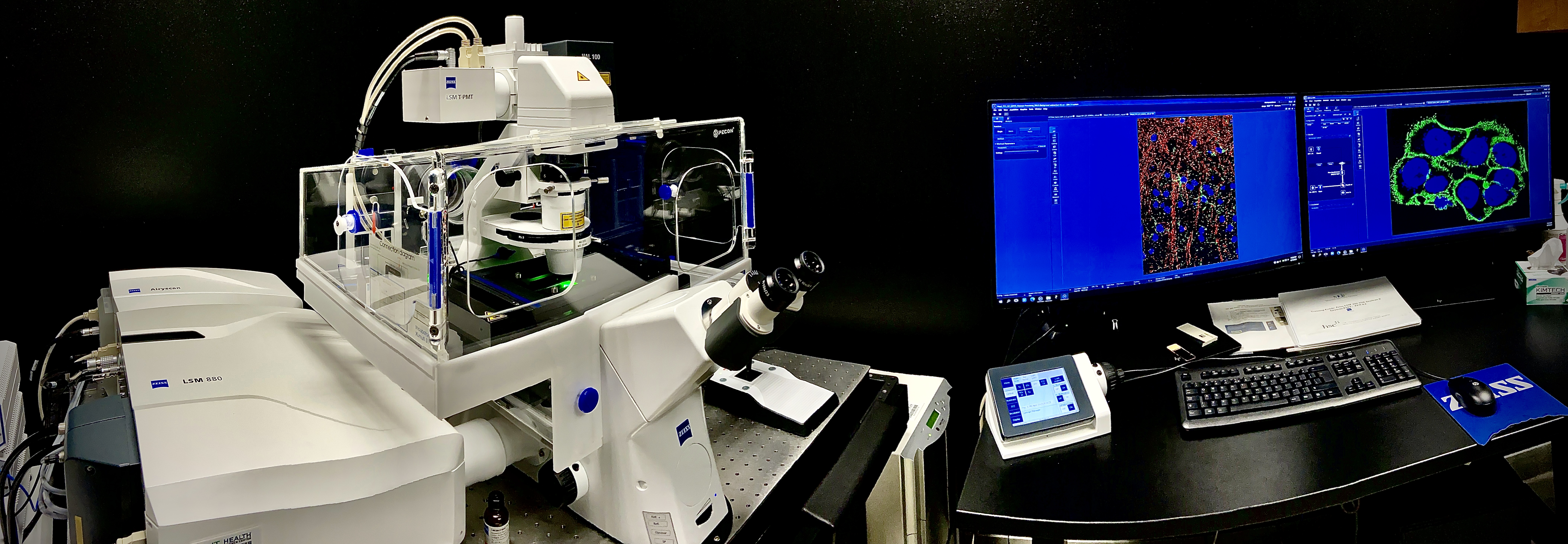
The Microscopy and Histology Core offers comprehensive support for histology and imaging experiments. We provide a range of services, including equipment training, consultation, and assistance with all aspects of the imaging workflow, from histology sample preparation to microscopy image acquisition and image analysis.
Our core facility is equipped with state-of-the-art microscopy systems and histology equipment to ensure high-quality sample preparation & imaging results.
The primary objective of this Core Facility is to cater to the research needs of the Health Science Center (HSC) and the broader scientific community in Fort Worth. We provide cutting-edge facilities that are open to faculty members, staff, and graduate students as well as external individuals and organizations enabling them to carry out their research effectively.
Microscopy Equipment
- Zeiss LSM 880 Confocal with AiryScan
- Zeiss Axioscan 7 (Slide Scanner)
- Keyence BZ-X800 All-in-one Fluorescence Microscope
- HP Z6 Image Analysis Workstation
Histology Equipment
- Tissue-Tek VIP AI Tissue Processor
- Tissue-Tek Cryo3 Flex Cryostat
- Leica UC7 Ultramicrotome
- Leica Microtome
- Leica Embedding station
Contact
Contact us for more details about equipment, services, and scheduling time in the Microscopy Core.
iLab
Log in to iLab to see a full list of equipment and schedule time in the Microscopy Core.
Per Hour Charges Based on User Type
| Internal | NEXT Lab | External |
||
| LSM 880 Inverted | Unassisted | $35.00 | NA | NA |
| Assisted | $65.00 | $96.00 | $192.00 | |
| Zeiss Axioscan 7 | Unassisted | $20.00 | NA | NA |
| Assisted | $40.00 | $60.00 | $192.00 | |
| Keyence BZ-X800 | Unassisted | $0.00 | NA | NA |
| Assisted | $30.00 | $45.00 | $90.00 | |
| Workstation | Unassisted | $10.00 | $15.00 | $30.00 |
Off-hours (7pm to 7am – Authorized Experienced Internal Users) pricing
| LSM 880 Inverted | Unassisted | $25.00 |
Trained users have 24/7 access to Microscopy Core. Technical assistance is available on weekdays from 8 a.m. to 5 p.m.
Training is mandatory for internal users who wish to use the microscopes on their own.
Training on confocal will cost $110 per person and include 3 hours of demonstration and 3 hours of hands-on training.
Training on Zeiss Axioscan 7(Slidescanner) can take from 2-4 hours and depends on the training objectives and costs same as assisted imaging ($40/Hr). Please contact Kishor.
Inverted Zeiss LSM 880 Airyscan Confocal Training
Training will generally be held on the 2nd and 4th Wednesday of each month at 1 p.m. Please contact Kishor.
Q: What is this about?
A: Opportunity to participate in a training session on the Inverted Zeiss LSM 880 AiryScan, Confocal Microscope (Super resolution image capture capability).
Q: When will the next sessions be held?
A: Generally held on Second and Fourth Wednesday every month at 1 p.m. Please contact Kishor Kunwar to schedule the training.
Q: How do I book my training?
A: Request Training through iLab.
- Login with your EUID and Password
- Go to Request Services tab
- Go to Request Initial (Training) section and Initiate Request
- Fill out the form, complete the payment information and submit
Q: How much will it cost?
A: $110 which will cover participation in ~2.5 hours of Demonstration Training and 3 hours of MCF staff monitored hands-on use imaging your own samples (schedule with MCF staff within two months of initial Demonstration training).
Q: What will the training format be?
A: Training is divided into two parts, demonstration, and hands-on use.
- Demonstration: In person: one session of ~2.5 hours.
- Hands-on use: In-person: one session lasting 3 hours or two sessions of 1.5 hours each.
Q: What if I am not comfortable to use the microscope on my own after initial training?
A: Please request further hands-on training. Within first two months after demonstration training, you will be charged $35/hour.
Training Format
Training will take place in at least two sessions, the initial Demonstration session followed by Hands-on Use.
- Demonstration: Core staff will operate the microscope and trainees will observe and interact with staff.
- Hands-on Use: After completing Demonstration, Trainees will schedule with MCF staff to operate the microscope with core staff guidance using their own samples.
Questions
- Contact Kishor Kunwar, Microscopy Specialist
- Contact Sharad Shrestha, Core Labs Director
Open-source image processing and analysis software
- Fiji : A distribution of ImageJ which includes many useful plugins contributed by the community.
- Icy : Used for a wide range of processing and analysis applications.
- CellProfiler : Used for high-throughput image analysis and data mining.
- KNIME : Used for high-throughput image analysis and data mining.
- QuPath : Used for digital pathology and whole slide image analysis.
- ilastik : Consists of powerful machine learning features needed to identify or classify challenging structures.
- napari : It is a fast, interactive, multi-dimensional image viewer for Python. It is designed for browsing, annotating, and analyzing large multi-dimensional images.
Bio-image processing and analysis learning resources
- Useful Bio-Image analysis YouTube videos.
- Quantifying microscopy images: Top 10 tips for image acquisition
- Quantitative Imaging for Colocalization Analysis (pdf)
- Avoiding Twisted Pixels: Ethical Guidelines for the Appropriate Use and Manipulation of Scienti?c Digital Images
Bio-imaging Networking
- The Center for Open Bioimage Analysis (COBA)
- The international network of cutting-edge bioimaging facilities and communities: (Exchange of Experience, Training, Shadowing, Working Groups)
Bio-imaging Sample Preparation
Zeiss LSM 880 Confocal with AiryScan and AiryScan Fast Mode
This system is based on a fully motorized Axio Observer 7 Inverted Microscope with Motorized Scanning Stage with Piezo Z and includes the following:
LSM Detectors (Spectral detection of 4 channels with two GaAsP detectors)
- 2 multi-anode and 1 GaAsP PMT
- Includes additional Airyscan GaAsP detector for super-resolution and Fast Mode
- Transmitted light detector T-PMT for detection of DIC, IR-DIC with laser illumination
Incubator Enclosure
Maintains user set CO2 level, temperature & humidity around the sample stage and objectives for live cell experiments.
Installed Lasers
- 405 nm Diode Laser 11.5 mW
- Argon Laser: 458/488/514 nm 15.4 mW
- 561 nm DPSS Laser 13 mW
- 633 nm HeNe Laser 2.3 mW
Objectives
- 5x/0.16 EC Plan-Neofluar 5x/0.16 WD=18.5
- 10x/.30 EC Plan-Neofluar 10x/0.30 WD=5.2
- 10X/0.5 W Plan-Apochromat 10x/0.5 WD=3.7mm
- 20x/0.8 Plan-Apochromat 20x/0.8 WD = 0.55
- 40x/1.1 W Corr FWD=0.62mm at CG=0.17mm
- 60X/0.7 Olympus LUC Plan FLN WD=1.5 – 2.2mm
- 63x/1.4 Oil Plan-Apochromat 63x/1.40 Oil
Zeiss Axioscan 7 (slidescanner)
- 100 slide capacity magazine plus Continuous Loading capability with auto sample detection and auto focus.
- High End Brightfield imaging with Axiocam 705 color camera and Fluorescent imaging with Axiocam 712 monochrome camera for high-resolution color acquisition
- High Performance Objectives: 5x, 10x/.45, 20x/0.8, 40x/.95 and circular polarizer set with 20x/0.5 POL objective
- Colibri 7 LED light source: (Excitation Wavelengths: 385nm, 430nm, 475nm, 555nm, 590nm, 630nm, 735nm) with continuous brightness adjustment for imaging of up to 7 channels
- High Efficiency Multi-band Filter: 110 and 112 for imaging of: DAPI, GFP, Cy3, mCherry, Cy5, Cy7, and similar dyes (Alexa 488, 555, 594, 647, 730)
- High Efficiency Single Pass Filter Sets: 38, 43, 50, and 96 for specific imaging of GFP, Cy3, Cy5 and similar dyes (Alexa 488, 555, 647)
- Premium HP Z6 Workstation: for large data handling with Windows 10 Enterprise
Zeiss LSM 880 Confocal with 2-Photon excitation laser
This system is based on a fully motorized upright Microscope with Motorized Scanning Stage includes the following:
LSM Detectors (Spectral detection of 4 channels with two GaAsP detectors)
- 2 multi-anode and 1 GaAsP PMT
- Transmitted light detector T-PMT for detection of DIC, IR-DIC with laser illumination
Installed Lasers
- 405 nm Diode Laser 11.5 mW
- Argon Laser: 458/488/514 nm 15.4 mW
- 561 nm DPSS Laser 13 mW
- 633 nm HeNe Laser 2.3 mW
- 690-1040nm tunable Mai-Tai/ Spectra-physics 2 photon laser
Objectives
- 10x/.30 EC Plan-Neofluar 10x/0.30 WD=5.2
- 10X/0.5 W Plan-Apochromat 10x/0.5 WD=3.7mm
- 20x/0.8 Plan-Apochromat 20x/0.8 WD = 0.55
- 20X/1 clarity dipping objective
- 40X/0.8 water dipping objective WD = 3.2mm
- 60X/0.7 Olympus LUC Plan FLN WD=1.5 – 2.2mm
Microscopy Core houses a Zeiss LSM confocal microscope, Keyence BZ-X800 fluorescence microscope and a data analysis station. Core provides access to state-of-the-art instruments for light microscopy imaging and software for image processing and analysis. A full-time staff provides training and consultation as well as assisted imaging on a fee-for-service basis.
Zeiss LSM 880 inverted confocal microscope with Airyscan super resolution capability is a primary microscope which is capable of simultaneous capture of four channel images using 4 laser lines which can produce 6 different laser wavelengths. It is equipped with 5 detectors, a GaAsP detector, an airyscan GaAsP detector, 2 multi anode PMT detectors and a T-PMT detector. The Airyscan feature can achieve 1.7X resolution (140nm XY, 400nm Z) and 4-8X sensitivity compared to standard confocal. Airyscan Fast is a unique feature that provides high resolution, sensitivity and speed up to 100fps that enables gentle live cell imaging with super resolution. This equipment is also equipped with definite focus, a piezo stage and a stage top live cell incubation system which maintains temperature, humidity, and CO2.
Keyence BZ-X800 fluorescence microscope is a fully motorized control system with advanced imaging and analysis functions. This inverted fluorescence microscope has a built-in darkroom and environment control system for live cell imaging. It is equipped with a high-sensitivity, cooled CCD camera that can be switched between monochrome and color imaging, and supports fluorescence, brightfield, and phase contrast imaging.
Confocal Microscope is connected with 10GB Ethernet cable to an image analysis workstation which is a high-performance computer with full feature Zen Black, Zen Blue and ImageJ/FIJI, Napari, and Cell profiler image analysis software installed.
Room housing the microscope has a CO2 incubator and a refrigerator to provide users with flexibility in maintaining their live cell or tissue sample during the facility use.
- How can I start accessing the Microscopy Core?
- Email Kishor.Kunwar@unthsc.edu to
- Schedule a training with Core staff, get trained and use microscopes by yourself.
- Schedule assisted imaging session with Core staff, who will coordinate with you to image your sample.
- Email Kishor.Kunwar@unthsc.edu to
- How can I get access to microscopy instruments after hours?
- Only trained users are authorized to operate instrument independently after hours.
- How can I get access to data analysis software?
- Schedule Imaging Workstation which is a HP Z6 high performance computer with software installed and located in RES111. Or you can use open source software on your own device.
- Who to contact if I have any question?
- Contact Kishor.Kunwar@UNTHSC.edu
Recommended Alexa Dyes for IHC/ICC
| Available Laser Excitation wavelength/Channel | Compatible Alexa Dyes (In order of compatibility with laser) |
| 405 | Alexa 405 | DAPI / Hoechst |
| 458 | Alexa 430 |
| 488 | Alexa 488 |
| 514 | Alexa 514 | Alexa 532 |
| 561 | Alexa 546 | Alexa 555 | Alexa 568 | Alexa 594 |
| 633 | Alexa 635 | Alexa 647 |
Live Cell Imaging tools
Chamber slides as well as cover-glass bottom plates can be used for confocal imaging.
Fixed cell ICC can be done by growing cells on coverglass and mounting the coverglass on the slide or growing cells on the slide (Chamberslides) and mounting with coverglass or any other plate or slides where live cell imaging can be done.
Live cell imaging will however require, cover glass chamber slide such as Lab-Tek II Part numbers 155409 (8 wells), 155382 (4 wells), 155379 (2 wells), and 155360 (1 well) or cover glass bottom plate MatTek Part number P06G-1.5-14-F (6 wells), P12G-1.5-14-F (12 wells), P24G-1.5-13-F (24 wells), Cover glass bottom dish MatTek Corporation 35 mm round dishes with #1.5 coverslip bottoms: Part number P35G-1.5-14-C.
Cover Glass
All our microscopes are tuned to image optimally with No. 1.5 (~170um) coverglass, always use the No. 1.5 coverglass.
- VWR Micro Cover Glasses, No. 1.5.
- Part number 48366-205 (18 x 18 mm), 48366-227 (22 x 22 mm), 48366-249 (25 x 25 mm).
- Warner Instruments , No. 1.5.
- Part numbers 64-0721(22 x 22 mm), 64-0716 (22 x 30 mm), 64-0717 (22 x 40 mm).
- For High resolution Airyscan imaging use: Warner Instruments High Tolerance #1.5 (0.17 ± 0.01 mm) Round Coverslips: Part numbers 64-0732 (12 mm), 64-0733 (15 mm), 64-0734 (18 mm), and 64-0735 (25 mm).
Mounting Medium
- ProLong Gold (Ideal for Alexa Fluor dyes), ProLong Diamond (Ideal for Alexa Fluor and traditional dyes, such as FITC and Cy3, and fluorescent proteins like GFP, RFP, and mCherry).
- For oil immersion objective imaging, use curing Prolong Glass Antifade Mountant (Which hardens after application) or non-curing SlowFade Glass Soft-set Antifade Mountant (Recommended for precise 3D measurements).
Mounting Media with DAPI is not recommended as it adds background to the fluorescence signal of your image.
Contributions to scientific output is an important metric for Core Labs, as it enables us to obtain financial and other support to ensure continuous high standards of operations to foster innovative research.
Users are encouraged to acknowledge Microscopy Core in presentations, posters, papers, and all other publications.
For example: “We acknowledge [name of staff] in the HSC Microscopy Core for training and assistance on [following]”.
When to acknowledge or provide co-authorship
- Include an acknowledgement any time the staff member or the Core provides services that support your research
- If a staff member has made a significant intellectual contribution beyond routine sample analysis, please consider co-authorship

